Microbiology of spoilage
Morphology, structure and growth of bacteria
Morphology
With a few exceptions, bacteria come in two types: cocci, which
are spherical, and rods, which are cylindrical. Cocci are 0.5 - 1µ in
diameter; rods are 0.3 - 1 µ in diameter and 1 - 10µ in length. 1
µ is 1/1000 mm. The size of cells can be measured directly by microscopic
observation or by the ability of cells to pass through a filter with pores of
known size.
The normal method of reproduction in bacteria is binary fission
in which a single cell divides into two identical daughter cells. It is often
found that the cells do not separate after division and the mass of cells that
results adopt characteristic patterns. If the planes of division are random the
pattern will resemble a bunch of grapes and is called staphylococci; divisions
in the same plane will result in a chain called streptococci.
Rod shaped cells can be regular cylinders or cigar shaped. They
are sometimes found in chains but are more often as single units. Some cells
contain a resistant spore which can be seen under the microscope; this will be
discussed in more detail later.
When bacteria are cultured on a solid nutrient, the colony which
is formed by the growing mass of cells is often of use in identifying the
bacterium; the addition of reagents and indicators can modify the appearance of
the colony and provide further information.
Structure
Microscopic examination of cells shows very little unless
special staining techniques are used. This is because the cell contents have a
refractive index which is similar to that of water which is the suspending
medium. In order to make a stained preparation, the cells must be spread thinly
on a glass slide and then fixed by gentle heating. Special dyes are then applied
to the smear and, after a suitable time, the excess is washed away leaving a
stained film of bacteria against a clear background. The dyes which are used
differ according to the chemical nature of the structure under study.
The nucleus contains the deoxyribonucleic acid (DNA) of the cell
and it is this which dictates the nature of the cell. In many cells, the nucleus
is a diffuse structure and can only be seen clearly during cell division when it
becomes thicker.
The ribosomes are the parts of the cell that translate the coded
information in the DNA into proteins, which are used to build the cell, and
enzymes, which control the biochemical processes which occur in the ceil.
The nucleus and the ribosomes are contained in the cytoplasm
which is bounded by the cytoplasmic membrane. The shape of the cell is governed
by the presence of a rigid cell wall, which may be protected by a slime layer.
Some cells are motile and they achieve this by means of a
whip-like flagellum. The flagellum arises from a basal granule in the cytoplasm
and protrudes through the cell wall. The position of the flagellum, or the
arrangement if there are more than one, is sometimes of use in identifying
bacteria.
The resistant spores referred to earlier are produced as a means
of surviving extremes in the environment. The spore is formed from only part of
the cell but always includes the nucleus with its DNA. When conditions are once
again favourable for growth the spore germinates and a new generation of cells
is produced. The spore which is dormant is protected by a tough spore coat and
by the fact that it has a very low water content. The position and size of a
spore within the cell is often of diagnostic use.
The chemical composition of the cell determines the way in which
it reacts to staining. In practice, the majority of cells fall into one of two
types. The first worker to discover this was a Dane, Gram, and this effect is
now called the Gram reaction in memory of him. He discovered that, using a
combination of dyes and decolourising solutions, cells would either stain blue
or red. We now know that this effect is determined by the chemistry of the cell
wall. The Gram reaction has proved to be a most valuable diagnostic tool.
Growth
When we talk about growth in bacterial terms, we usually refer
to the increase in numbers of cells and not the enlargement of individual cells.
It is a fairly simple procedure to count the numbers of cells in a liquid
nutrient medium and, from this data, we can produce a growth curve for the
population.
In most spoilage situations, there is a limited supply of
nutrients and the growth curve under these conditions usually takes the form
shown in Figure 15. For reasons that need not concern us here, the numbers of
cells is plotted as the logarithm of the actual number counted against time.
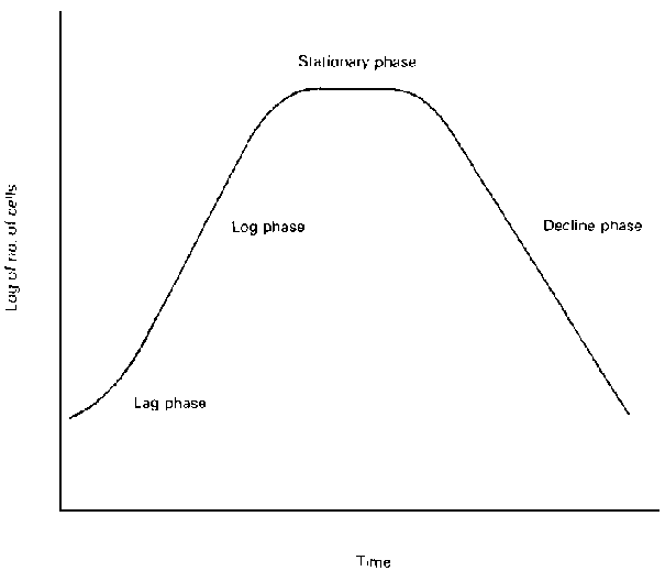
Figure 15 - Typical growth curve
At the start of the curve, the cells are adapting to the new
environment and the rate of division is so slow that it does not keep pace with
the number of cells which are dying. This tends to give a straight horizontal
line or a slight downward curve. This is called the lag phase,
At the end of the lag phase, the cells begin to divide more
rapidly and the total number of cells in the culture rises. At best, the cells
will reproduce logarithmically which on our graph is shown by a straight line,
the slope of which is a measure of the rate of growth. This is called the log
phase.
After a time, the nutrients become exhausted and there may be a
build up of toxic waste products. These and other factors lead to a reduction in
the rate of cell division and a consequent flattening of the growth curve. At
this point cell division and death are equal and this is called the stationary
phase.
Following the stationary phase, the rate of division falls even
more and there is an increase in cell death which leads to a logarithmic decline
phase. Introduction of fresh nutrients, or the transfer of some cells to a new
source of nutrient, will result in a repeat of the cycle.
From experiments of this type, it is possible to calculate the
time taken for a newly formed cell to mature and reproduce itself: this is
called the generation time. Different bacteria, and even the same bacteria under
different conditions, have different generation times. Many preservation
techniques control spoilage by inducing a prolongation of the generation time.
Microbial classification
Bacteria, like most living things, have been sorted into groups
which have similarities. The distinguishing features are normally biochemical
and morphological. The system as a whole need not concern us except to point out
that in referring to a particular organism use is made of both the generic and
specific names. Thus Staphylococcus aureus is usually abbreviated to S. aureus.
Yeasts and moulds
Yeasts are usually ellipsoidal and are about 6µ by 3µ
in size. They reproduce by budding although some do divide in a manner similar
to bacteria. Moulds consist of cylindrical filaments called hyphae which form a
mass known as the mycelium. The hyphae may or may not have crosswalls. Moulds
reproduce both sexually and asexually, in both cases a spore (or spores) is
formed which can germinate to form a new mycelium. Fragments of hyphae will also
produce a new mycelium if the environment is suitable.
Yeasts are not important as far as seafoods are concerned but
moulds, due to their ability to grow where water is limited, can be a problem on
smoked or dried fish. Where water is abundant, the bacteria grow so much more
rapidly that moulds are only of secondary importance.
Culturing bacteria
In order to study bacteria, or for that matter moulds, they must
be separated from the sample and grown on an artificial food source. The
composition of the growth medium must be optimal for the particular organism and
this means that the exact requirements of each organism must be known.
Fortunately, most of this work has been done for the commonly isolated organisms
and we have only to prepare the medium as laid down in the standard texts on the
subject.
The temperature at which the medium is incubated must suit the
organism and there are three groups: psychrotrophs which grow at 0 - 5°C
but have higher optimum temperatures; mesophiles which favour temperatures
between 10 and 45°C; and thermophiles which grow at 80°C. At chill
temperatures, the psychrotrophs are important spoilage organisms whereas
organisms of public health significance are almost always mesophiles.
When an organism is growing on a suitable medium, it becomes
easy to test its various biochemical properties and study the shape and size of
the colonies. The ability to ferment certain sugars or utilise unusual carbon or
nitrogen sources can be determined and is useful in identifying unknown
cultures.
Bacteria may be grown in tubes of liquid media, or on slopes or
plates of media solidified with agar. Cotton wool plugs are the traditional
method of sealing tubes and bottles of media but screw caps have the advantage
that there is a reduction in evaporation which means that media have a longer
shelf
life.
