| Minor Surgical Procedures in Remote Areas (MSF, 1989, 172 p.) | ||||
| Chapter 2: Infection of soft tissues | ||||
| Abscess | ||||
| Treatment of tropical myositis | ||||
| Infections of the hand and fingers | ||||
|
| |||||||||||||||||||||||||
Abscess anesthesia of abscesses and tropical myositis
Anesthesia of an abscess with lidocaine is not very effective. It is not possible to anesthetise the whole cavity and furthermore, the needle may spread the infection.
General anesthesia is preferable: ketamine IM, at the rate of 5mg/kg for exemple.
For superficial abscesses, use ethylene chloride: anesthetise the area and incise immediately (the anesthetic freezes the skin by evaporation). abscess
A collection of pus in the soft tissues. An abscess cavity is not accessible to antibiotics. Treatment is thus surgical only.
Indications
Incision and drainage (I & D) should be performed once the abscess is "ripe" i.e. fluctuant upon gentle palpation.
Material
· Sterile scalpel blade and handle
· Surgical gloves
· Plain curved forceps (Kelly forceps)
· Sterile corrugated drain
· Antiseptic s, e.g. chlorhexidine (+ cetrimide) solution (see table page 7).
Technique
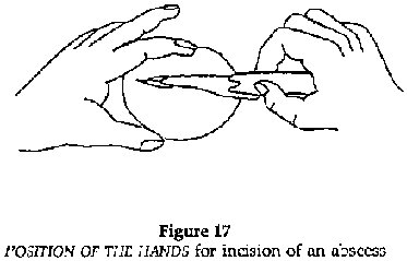
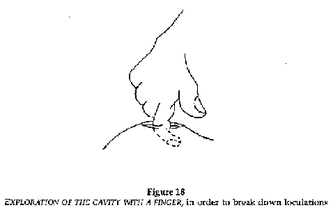
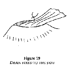
Figures 17,18 and 19
· Scalpel: the correct way to hold a scalpel is between the thumb and forefinger with the handle resting against the palm. Il should not be held as one holds a pen. The plane of the scalpel blade should be perpendicular to the plane of the skin.
· Incision: the free hand immobilizes the wall of the abscess between thumb and forefinger. Incise in the long axis of the abscess with a single stroke to breach the skin. The incision should be long enough to allow insertion of an exploring finger.
· Take care not to incise too deeply if the abscess overlies major blood vessels (the carotid, axillary, humeral, femoral and popliteal regions). After breaching the skin, blunt dissect down to the cavity using Kelly's forceps.
· Explore the cavity with the forefinger, breaking any loculating adhesions and evacuating the pus. Abundant lavage of the cavity using a syringe filled with chlorhexidine (+ cetrimide) (see table page 7).
· Insert a drain, if possible fixing it with a single suture at the edge of the incision. The drain is withdrawn progressively then removed altogether after 3 to 5 days.
FIGURE
FIGURE
FIGURE
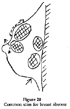
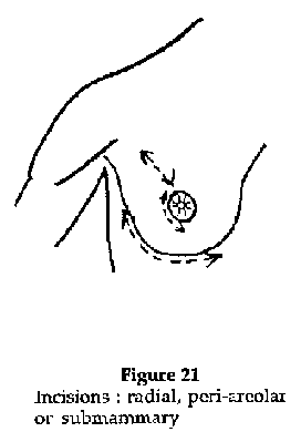
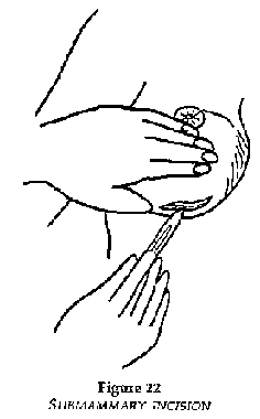
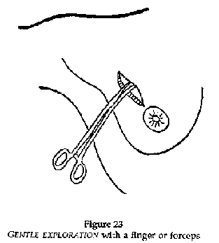
Breast abscess
Figure 20
Usually the abscess is superficial but deep ones, when they occur, are more difficult to diagnose and treat.
Early in the infection, before the infection loculates (mastitis), it is possible to treat medically: antibiotics, anti-inflammatories, hot compresses, constricting bandage to reduce lactation in the affected breast and expression of milk to avoid engorgement.
Fluctuation occurs early in superficial infections and later for deeper infections.
Material
Same material as for other abscesses.
Technique
· Radial incision for superficial abscesses, peri-areolar for abscesses near the nipple (Figure 21).
· Submammary incision for deep abscesses (Figure 22).
· Gentle exploration with finger or forceps to break down loculations (Figure 23).
· Abundant lavage with chlorhexidine (+ cetrlmide) solution (see table page 7).
· Insertion of a large drain.
FIGURE
FIGURE
FIGURE
FIGURE
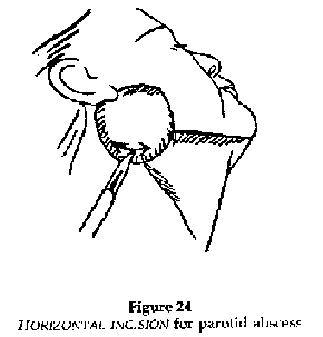
Parotid abscess
The incision is a risk to the branches of the facial nerve. Consequently, the incision should be over the caudal part of the abscess and parallel to the lower border of the maxilla.
FIGURE
Anesthesia
Treatment of myositis
Example: myositis of the quadriceps
Infection and eventually abscess formation within muscle sheaths.
At the stars of infection, when the muscle is swollen, hot and painful, medical treatment may prevent abscess formation: immobilize, give antiinflammatory medication (Indomethacin (PO): 75 mg/d in 3 divided doses x 5 days), and antibiotics (ampicillin (PO): 4 g/d in 4 divided doses x 7 days for an adult).
Indications
When the swelling becomes fluctuant conduct an exploratory puncture with a largebore needle which, if the diagnosis is correct, will produce thick pus (Figure 25).
Material
The same as for an abscess.
Technique
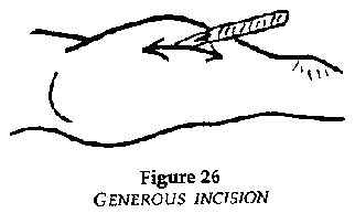
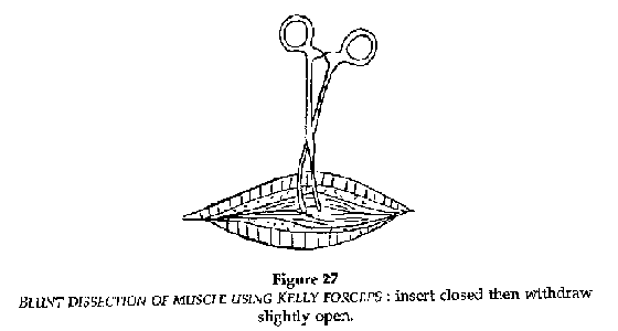
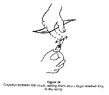
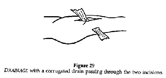
(Figures 25, 26, 27, 28, 29)
· Generous skin incision, avoiding underlying neurovascular bundles, and incision of the fascia and muscle sheath.
· Blunt dissection with Kelly forceps or rounded scissors; push the closed instruments into the muscle as far as the abscess cavity and gently open them and withdraw.
· Explore with a finger to break the adhesions and evacuate the pus.
· Lavage with chlorhexldine (+ cetrimideJ (see table page 7).
· If possible, place a counter incision on the skin near the edge of the abscess, cutting down on a finger that is inserted deep in the cavity.
· Pass a strip of corrugated drain between the two incisions.
· Fix the drain with a suture to the edge of the incision and withdraw around the 5th day.

FIGURE
FIGURE
FIGURE
FIGURE
FIGURE
Myositis of the psoas muscle
If the abscess is on the right side, the clinical signs are the same as for appendicitis with pain in the right iliac area.
Treatment requires surgical intervention as far as the peritoneum. This is the only way to make the diagnosis and consequently the operator must be able to carry out an appendicectomy if required.
In the case of a psoas abscess, once the peritoneum has been reached, it should be separated, not opened and the psoas should appear behind. Open the sheath (blunt dissection with rounded scissors; insert closed then withdraw slightly open) and release the pus. Clean and drain as with other abscesses.
Paronychia
Infection of a finger is a paronychia.
Indications
Antibiotics, frequently prescribed during the acute phase, retard the collection of pus and consequently drainage. Patients are seen most often during the acute phase.
Superficial paronychia
Material
Scalpel, pair of rounded scissors, non toothed dissecting forceps, gloves, sterilized drapes, and equipment for a local anesthetic.
Anesthesia
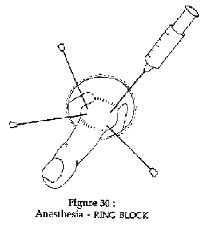
Figure 30
Ring block with lidocaine 1%, or anesthetise the skin with ethylene chloride.
Technique
· Paronychia at the edge of the nail
Lift and separate the edge of the nail with a scalpel at the point of oedema and inflammation; pus will escape. Apply daily dressings.
· Subungual paronychia
The nail is elevated by pus between the nail and the tip of the finger. Cut a triangle between the nail bed and the pulp and excise the pus. Apply daily dressings.
· Peri-ungual paronychia
The edge of the nail and the base are separated by the abscess. Incise the skin at the nail base and continue along the nail edge separating the skin from the nail base. Lift part of the nail, that covering the abscess, but leave the part which protects the ungual phalanx and assists healing. Apply daily moist dressings.
· Paronychia on the dorsum of the finger
Almost always occurs on the index finger. Incise with a scalpel and dress daily. Give antibiotics (e.g. ampicillin: 4 g/d for an adult, in 4 divided doses for a minimum of 5 days) to avoid the spread of infection and risk to the extensor tendon.
Subcutaneous paronychia
· Paronychia of the finger pulp: deep infection
One or two lateral incisions, on the dorsal and ventral surfaces of the finger, avoiding the vascular/nerve bundles. Insert a small drain for two days. Apply daily moist dressings.
· Paronychia of the two first phalanges
One or two lateral and dorsal incisions at the superior border of the two commissures, avoiding the vascular/nerve bundles. Apply daily moist dressings.
Osteitis of the finger
Suspect a persistent fistula or even a fistula communicating with bone when an incision demonstrates the presence of a sequestration. Bony excision must be minimal; it is necessary to limit excision to the sequestered bone and to avoid amputation.
Prescribe a large dose and a long course of antibiotics (ampicillin: 4g/day for an adult, for a minimum of 4 weeks).
Immobilise in flexion with a splint.
Tenosynovitis of the finger
Collection of pus in the sheath of the flexor tendon of the finger.
Diagnostic
The finger is bent with pain running along its length.
Material
Surgical kit
Anesthesia
General (Ketamine) or local/regional.
Technique
· First make a vertical palmer incision of 2.5 cm at the head of the metacarpal.
· Shield and protect the vascular/nerve bundles.
· Identify the cul-de-sac of the dilated sheath and incise on both sides. Clean with chlorhexidine (+ cetrimide) solution (see table 1).
· Place closed forceps into the incision through to the skin on the opposite side and make a counter incision with a scalpel.
· Pass a drain between the two incisions. This drain remains until the swelling disappears.
· Immobilise with a splint and prescribe antibiotics (e.g. ampicillin : 4 g/d for an adult in 4 divided doses for at least 8 days).
· Apply daily dressings.