Back to Home
Page of CD3WD Project or Back to list of CD3WD Publications
 |  |  | Where There Is No Doctor - A Village Health Care Handbook (Hesperian Foundation, 1993, 516 p.) |  |  | Chapter 16 - THE EYES | |  | (introduction...) | | | Danger Signs | | | Injuries to the Eye | | | How to Remove a Speck of Dirt from the Eye | | | Chemical Burns of the Eye | | | Red, Painful Eyes - Different Causes | | | ‘Pink Eye’ (Conjunctivitis) | | | Trachoma | | | Infected Eyes in Newborn Babies (Neonatal Conjunctivitis) | | | Iritis (Inflammation of the Iris) | | | Glaucoma | | | Infection of the Tear Sac (Dacryocystitis) | | | Trouble Seeing Clearly | | | Cross-Eyes and Wandering Eyes | | | Sty (Hordeolum) | | | Pterygium | | | A Scrape, Ulcer, or Scar on the Cornea | | | Bleeding in the White of the Eye | | | Bleeding behind the Cornea (Hyphema) | | | Pus Behind the Cornea (Hypopyon) | | | Cataract | | | Night Blindness and Xerophthalmia (Vitamin A Deficiency) | | | Spots or 'Flies' before the Eyes (Mouches Volantes) | | | Double Vision | | | River Blindness (Onchocerciasis) |
|
Where There Is No Doctor - A Village Health Care Handbook (Hesperian Foundation, 1993, 516 p.)
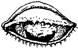
Chapter 16 - THE EYES
Figure
Danger Signs
The eyes are delicate and need good care. Get medical help fast
when any of the following danger signs occurs:
1. Any injury that cuts or ruptures (goes through)
the eyeball.
2. Painful, grayish spot on the cornea, with redness around the
cornea (corneal ulcer).
3. Great pain inside the eye (possibly iritis or glaucoma).
4. A big difference in the size of the pupils when there is pain
in the eye or the head.
A big difference in the size of
the pupils may come from brain damage, stroke, injury to the eye, glaucoma, or
iritis. (A small difference is normal in some people.)
5. Blood behind the cornea inside the eyeball.
Figure
6. If vision begins to fail in one or both eyes.
7. Any eye infection or inflammation that does not get better
after 5 or 6 days of treatment with an antibiotic eye
ointment.
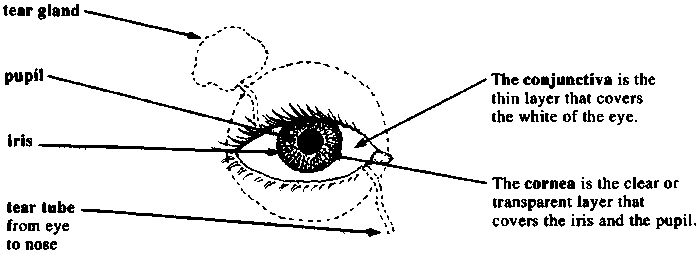
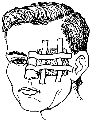
Injuries to the Eye
All injuries to the eyeball must be considered dangerous, for
they may cause blindness.
Even small cuts on the cornea (the transparent layer
covering the pupil and iris) may get infected and harm the vision if not cared
for correctly.
If a wound to the eyeball is so deep that it reaches the black
layer beneath the outer white layer, this is especially dangerous.
If a blunt injury (as with a fist) causes the eyeball to fill
with blood, the eye is in danger. Danger is especially great if pain suddenly
gets much worse after a few days, for this is probably acute glaucoma.
Treatment:
· If the person
still sees well with the injured eye, put an antibiotic eye ointment in the eye
and cover it with a soft, thick bandage. If the eye is riot better in a day or
two, get medical help.
· If the person cannot see well
with the injured eye, if the wound is deep, or if there is blood inside the eye
behind the cornea, cover the eye with a clean bandage and go for medical help at
once. Do not press on the eye.
· Do not try to remove
thorns or splinters that are tightly stuck in the eyeball. Get medical
help.
Figure
How to Remove a Speck of Dirt from the Eye
Have the person close her eyes and look to the left, the right,
up and down. Then, while you hold her eye open, have her look up and then down.
This will make the eye produce more tears and the dirt often comes out by
itself.
Or, you can try to remove the bit of dirt or sand by flooding
the eye with clean water or by using the corner of a clean cloth or some moist
cotton. If the particle of dirt is under the upper lid, look for it by turning
the lid up over a thin stick. The person should look down while you do this:
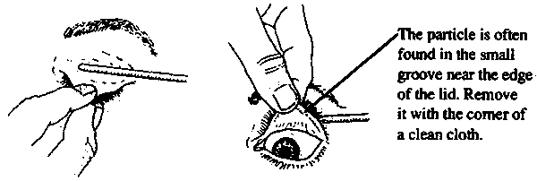
Figure
If you cannot get the particle out easily, use an antibiotic eye
ointment, cover the eye with a bandage, and go for medical
help.
Chemical Burns of the Eye
Figure
Battery acid, lye, gasoline, or a pesticide that gets into the
eye can be dangerous. Hold open the eye. Immediately flood the eye with
clean, cool water. Keep flooding for 30 minutes, or until it stops hurting.
Do not let the water get into the other
eye.
Red, Painful Eyes - Different Causes
Many different problems cause red, painful eyes. Correct
treatment often depends on finding the cause, so be sure to check carefully for
signs of each possibility. This chart may help you find the cause:
|
foreign matter (bit of dirt, etc.) in the eye |
usually affects one eye only;
redness and pain variable |
|
burns or harmful liquids |
one or both eyes;
redness and pain variable |
|
pink eye' (conjunctivitis)
hay fever (allergic conjunctivitis)
trachoma measles |
usually both eyes (may start or be worse in one)
usually reddest at outer edge
Figure
'burning' pain, usually mild |
|
acute glaucoma
iritis
scratch or ulcer on the cornea |
usually one eye only;
reddest next to the cornea
Figure
pain often great |
‘Pink Eye’ (Conjunctivitis)
This infection causes redness, pus, and mild “burning”
in one or both eyes. Lids often stick together after sleep. It is especially
common in children.
Treatment:
First clean pus from the eyes with a clean cloth moistened with
boiled water. Then put in antibiotic eye ointment. Pull down the lower lid and
put a little bit of ointment inside, like this: Putting ointment outside
the eye does no good.
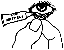
Figure
CAUTION: Do not touch the tube against the eye.
Prevention:
Most conjunctivitis is very contagious. The infection is easily
spread from one person to another. Do not let a child with pink eye play or
sleep with others, or use the same towel. Wash hands after touching
eyes.
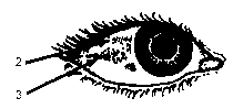
Trachoma
Trachoma is a chronic infection that slowly gets worse. It may
last for months or many years. If not treated early, it sometimes causes
blindness. It is spread by touch or by flies, and is most common where people
live in poor, crowded conditions.
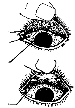
Figure
Signs:
· Trachoma begins
with red, watery eyes, like ordinary conjunctivitis.
· After a month or more, small,
pinkish gray lumps, called follicles, form inside the upper eyelids. (1)
· The white of the eye is a
little red. (2)
· After a few months, if you
look very carefully, or with a magnifying glass, you may see that the top edge
of the cornea looks grayish, because it has many tiny new blood vessels in it
(pannus). (3)
· The combination of both
follicles and pannus is almost certainly trachoma.
· After several years, the
follicles begin to disappear, leaving whitish scars. (4)
These scars make the eyelids thick
and may keep them from opening or closing all the way.
Or the scarring may pull the
eyelashes down into the eye, scratching the cornea and causing blindness.
Treatment of trachoma:
Put 1% tetracycline eye ointment inside the eye 3 times each
day, or 3% tetracycline eye ointment 1 time each day. Do this for 30 days. For a
complete cure, also take tetracycline or a sulfonamide by mouth for 2 to 3
weeks.
Prevention:
Early and complete treatment of trachoma helps prevent its
spread to others. All persons living with someone who has trachoma, especially
children, should have their eyes examined often and if signs appear, they should
be treated early. Washing the face every day can help prevent trachoma. Also, it
is very important to follow the Guidelines of Cleanliness, explained in Chapter
12.
|
Cleanliness helps prevent trachoma. |
Infected Eyes in Newborn Babies (Neonatal Conjunctivitis)
Figure
In the first 2 days of life, if a newborn baby's eyes get red,
swell, and have a lot of pus in them, this is probably gonorrhea. It must
be treated at once to prevent the baby from going blind. If the eye
infection begins between 1 and 3 weeks after birth, she may have
chlamydia. The baby has picked up one or both of these diseases from the
mother at birth.
Treatment for gonorrhea:
· Give one injection
of 50 to 75 mg. of kanamycin. Or, penicillin sometimes works: inject 200,000
units of crystalline penicillin twice a day for 3 days. Or, try 1/2 teaspoon of
co-trimoxazole syrup by mouth 2 times a day for a week. (If available, one
injection of 125 mg. of ceftriaxone is the best treatment.)
· Also use tetracycline eye
ointment: put a little in the baby's eyes every hour for the first day, and then
3 times a day for 2 weeks.
Treatment for chlamydia: Treat with tetracycline eye
ointment as described above. Also give erythromycin syrup by mouth, 30 mg. 4
times a day for 2 weeks. (This will treat pneumonia, which often affects babies
with chlamydia.)
Prevention:
All babies' eyes should be protected against gonorrhea
and chlamydia, especially the eyes of babies whose mothers may have these
diseases or whose fathers have pain when passing urine. (Mothers may have
gonorrhea or chlamydia without knowing it.)
Put a drop of 1% silver nitrate solution once only in
each eye at birth. Or, put a little 1% tetracycline eye ointment, or .5%
erythromycin eye ointment, in each eye at birth.
If a baby develops gonorrhea or chlamydia of the eyes, both
parents should be treated for these
infections.
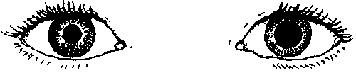
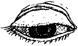
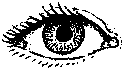
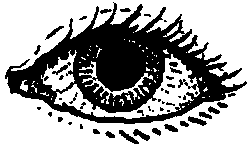
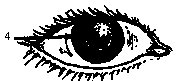
Iritis (Inflammation of the Iris)
Signs:
NORMAL EYE
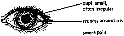
EYE WITH IRITIS
Iritis usually happens in one eye only. Pain may begin suddenly
or gradually. The eye waters a lot. It hurts more in bright light. The eyeball
hurts when you touch it. There is no pus as with conjunctivitis. Vision is
usually blurred.
This is an emergency. Antibiotic ointments do not
help. Get medical
help.
Glaucoma
This dangerous disease is the result of too much pressure in the
eye. It usually begins after the age of 40 and is a common cause of blindness.
To prevent blindness, it is important to recognize the signs of glaucoma and
get medical help fast.
There are 2 forms of glaucoma.
ACUTE GLAUCOMA:
This starts suddenly with a headache or severe pain in the eye.
The eye becomes red, the vision blurred. The eyeball feels hard to the touch,
like a marble. There may be vomiting. The pupil of the bad eye is bigger than
that of the good eye.
normal
glaucoma
If not treated very soon, acute glaucoma will cause blindness
within a few days. Surgery is often needed. Get medical help fast.
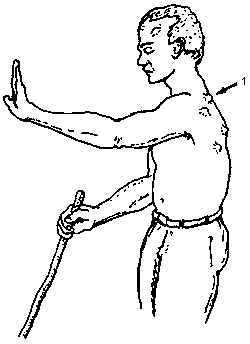
CHRONIC GLAUCOMA:
The pressure in the eye rises slowly. Usually there is no pain.
Vision is lost slowly, starting from the side, and often the person does not
notice the loss. Testing the side vision may help detect the disease.
Have the person cover one eye, and with the other look at an
object straight ahead of him. Note when he can first see moving fingers coming
from behind on each side of the head.
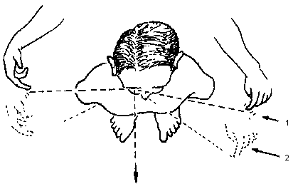
TEST FOR GLAUCOMA
Normally fingers are first seen here. (1)
In glaucoma, finger movement is first seen more toward the
front. (2)
If discovered early, treatment with special eyedrops
(pilocarpine) may prevent blindness. Dosage should be determined by a doctor or
health worker who can measure the eye pressure periodically. Drops must be used
for the rest of one's life. When possible, eye surgery is usually the surest
form of treatment.
Prevention:
Persons who are over 40 years old or have relatives with
glaucoma should try to have their eye pressure checked once a
year.
Infection of the Tear Sac (Dacryocystitis)
Signs:
Redness, pain, and swelling beneath the eye, next to the nose.
The eye waters a lot. A drop of pus may appear in the corner of the eye when the
swelling is gently pressed. (1)
Figure
Treatment:
· Apply hot
compresses.
· Put antibiotic eye drops or
ointment in the eye.
· Take
penicillin.
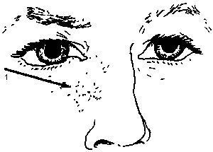
Trouble Seeing Clearly
Children who have trouble seeing clearly or who get
headaches or eye pain when they read may need glasses. Have their eyes examined.
In older persons, it is normal that, with passing years,
it becomes more difficult to see close things clearly. Reading glasses often
help. Pick glasses that let you see clearly about 40 cm. (15 inches) away from
your eyes. If glasses do not help, see an eye doctor.
Figure
Cross-Eyes and Wandering Eyes
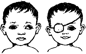
Figure
If the eye sometimes wanders like this, but at other times looks
ahead normally, usually you need not worry. The eye will grow straighter in
time. But if the eye is always turned the wrong way, and if the child is not
treated at a very early age, she may never see well with that eye. See an eye
doctor as soon as possible to find out if patching of the good eye, surgery, or
special glasses might help.
Surgery done at a later age can usually straighten the eye and
improve the child's appearance, but it will not help the weak eye see better.
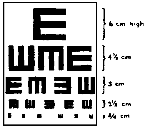
IMPORTANT: The eyesight of every child should be
checked as early as possible (best around 4 years). You can use an 'E' chart
(see Helping Health Workers Learn). Test each eye separately to discover
any problem that affects only one eye, eye. If sight is poor in one or both
eyes, see an eye doctor.
Figure
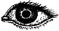
Sty (Hordeolum)
Figure
A red, swollen lump on the eyelid, usually near its edge. To
treat, apply warm, moist compresses with a little salt in the water. Use of an
antibiotic eye ointment 3 times a day will help prevent more sties from
occurring.
Pterygium
Figure
A fleshy thickening on the eye surface that slowly grows out
from the edge of the white part of the eye near the nose and onto the cornea;
caused in part by sunlight, wind, and dust. Dark glasses may help calm
irritation and slow the growth of a pterygium. It should, be removed by surgery
before it reaches the pupil. Unfortunately, after surgery a pterygium often
grows back again.
Folk treatments using powdered shells do more harm than good; To
help calm itching and burning you can try using cold compresses. Or use eye
drops of camomile (well boiled, then cooled, and without
sugar).
A Scrape, Ulcer, or Scar on the Cornea
When the very thin, delicate surface of the cornea has been
scraped, or damaged by infection, a painful corneal ulcer may result. If
you look hard in a good light, you may see a grayish or less shiny patch on the
surface of the cornea.
Figure
If not well cared for, a corneal ulcer can cause blindness.
Apply antibiotic eye ointment, 4 times a day for 7 days, give penicillin, and
cover the eye with a patch. If the eye is not better in 2 days, get medical
help.
A corneal scar is a painless, white patch on the cornea.
It may result from a healed corneal ulcer, burn, or other injury to the eye. If
both eyes are blind but the person still sees light, surgery (corneal
transplant) to one eye may return its sight. But this is expensive. If one eye
is scarred, but sight is good in the other, avoid surgery. Take care to protect
the good eye from injury.
Figure
Bleeding in the White of the Eye
Figure
A painless, blood-red patch in the white part of the eye
occasionally appears after lifting something heavy, coughing hard (as with
whooping cough), or being hit on the eye. The condition results from the
bursting of a small vessel. It is harmless, like a bruise, and will slowly
disappear without treatment in about 2 weeks.
Small red patches are common on the eyes of newborn babies. No
treatment is
needed.
Bleeding behind the Cornea (Hyphema)
Figure
Blood behind the cornea is a danger sign. It usually results
from an injury to the eye with a blunt object, like a fist. If there is pain and
loss of sight, refer the person to an eye specialist immediately. If the pain is
mild and there is not loss of sight, patch both eyes and keep the person at rest
in bed for several days. If after a few days the pain becomes much worse, there
is probably hardening of the eye (glaucoma). Take the person to an eye doctor
at
once.
Pus Behind the Cornea (Hypopyon)
Figure
Pus behind the cornea is a sign of severe inflammation.
It is sometimes seen with corneal ulcers and is a sign that the eye is in
danger. Give penicillin and get medical help at once. If the ulcer is treated
correctly, the hypopyon will often clear up by
itself.
Cataract
Figure
The lens of the eye, behind the pupil, becomes cloudy making-the
pupil look gray or white when you shine a light into it. Cataract is common in
older persons, but also occurs, rarely, in babies. If a blind person with
cataracts can still tell light from dark and notice movement, surgery may let
him see again. However, he will need strong glasses afterward, which take time
to get used to. Medicines do not help cataracts. (Now sometimes during surgery
an artificial lens is put inside the eye - so that strong eyeglasses are not
needed.)
Night Blindness and Xerophthalmia (Vitamin A Deficiency)
This eye disease is most common in children between 1 and 5
years of age. It comes from not eating enough foods with vitamin A. If not
recognized and treated early, it can make the child blind.
Signs:
· At first, the
child may have night blindness. He cannot see as well in the dark as
other people can. (1)
Figure
· Later, he develops dry
eyes (xerophthalmia). The white of the eyes loses its shine and begins to
wrinkle. (2)
· Patches of little gray bubbles
(Bitot's spots) may form in the eyes. (3)
Figure
· As the disease gets worse, the
cornea also becomes dry and dull, and may develop little pits. (4)
Figure
· Then the cornea may quickly
grow soft, bulge, or even burst. Usually there is no pain. Blindness may
result from infection, scarring, or other damage. (5)
Figure
· Xerophthalmia often begins, or
gets worse, when a child is sick with another illness like diarrhea, whooping
cough, tuberculosis, or measles. Examine the eyes of all sick and underweight
children. Open the child's eyes and look for signs of vitamin A
deficiency.
Prevention and treatment:
Xerophthalmia can easily be prevented by eating foods that have
vitamin A. Do the following:
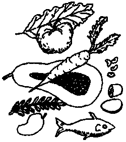
Figure
· Breast feed the baby - up to 2
years, if possible.
· After the first 6
months, begin giving the child foods rich in vitamin A, such as dark green leafy
vegetables, and yellow or orange fruits and vegetables such as papaya (paw paw),
mango, and squash. Whole milk, eggs, and liver are also rich in vitamin
A.
· If the child is
not likely to get these foods, or if he is developing signs of night blindness
or xerophthalmia, give him vitamin A, 200,000 units (60 mg. retinol, in capsule
or liquid) once every 6 months. Babies under 1 year of age should get 100,000
units.
· If the condition is already
fairly severe, give the child 200,000 units of vitamin A the first day, 200,000
units the second day, and 200,000 units 14 days later. Babies under 1 year old
should get half that amount (100,000 units).
· In communities where
xerophthalmia is common, give 200,000 units of vitamin A once every 6 months to
women who are breast feeding, and also to pregnant women during the second half
of their pregnancy.
WARNINGS: Too much vitamin A is poisonous. Do not
give more than the amounts advised here.
If the condition of the child's eye is severe, with a dull,
pitted, or bulging cornea, get medical help. The child's eye should be bandaged,
and he should receive vitamin A at once, preferably an injection of 100,000
units.
|
Dark green leafy vegetables, and yellow or orange fruits
and vegetables, help prevent blindness in children. |
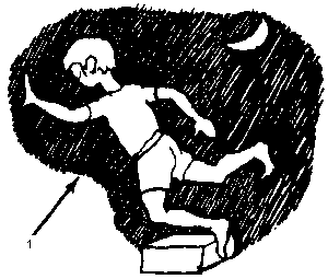
Spots or 'Flies' before the Eyes (Mouches Volantes)
Sometimes older persons complain of small moving spots when they
look at' a bright surface (wall, sky). The spots move when the eyes move and
look like tiny flies. These spots are usually harmless and need no treatment.
But if they appear suddenly in large numbers and vision begins to fail from one
side, this could be a medical emergency (detached retina). Seek medical help
at
once.
Double Vision
Figure
Seeing double can have many causes.
If double vision comes suddenly, is chronic, or gradually gets
worse, it is probably a sign of a serious problem. Seek medical help.
If double vision occurs only from time to time, it may be a sign
of weakness or exhaustion, perhaps from malnutrition. Read Chapter 11 on good
nutrition and try to eat as well as possible. If sight does not improve, get
medical
help.
River Blindness (Onchocerciasis)
This disease is common in many parts of Africa and certain areas
of southern Mexico, Central America, and northern South America. The infection
is caused by tiny worms that are carried from person to person by small,
hump-backed flies or gnats known as black flies (simulids).
BLACK FLY
The worms are 'injected' into a person when an infected black
fly bites him.
Signs of river blindness:
· Several months
after a black fly bites and the worms enter the body, lumps begin to form under
the skin. In the Americas the lumps are most common on the head and upper body;
in Africa on the chest, the lower body, and thighs. Often there are no more than
3 to 6 lumps. They grow slowly to a size of 2 to 3 cm. across. They are usually
painless. (1)
Figure
· There may be itching when the
baby worms are spreading.
· Pains in the back, shoulder or
hip joints, or 'general pains all over'.
· Enlargement of the lymph nodes
in the groin.
· Thickening of the skin on the
back or belly, with big pores like the skin of an orange. To see this, look at
the skin with light shining across it from one side.
· If the disease is not treated,
the skin gradually becomes more wrinkled, like an old man's. White spots and
patches may appear on the front of the lower legs. A dry rash may appear on the
lower limbs and trunk.
· Eye problems often lead to
blindness. First there may be redness and tears, then signs of iritis. The
cornea becomes dull and pitted as in xerophthalmia. Finally, sight is lost
because of corneal scarring, cataract, glaucoma, or other problems.
Treatment of river blindness:
Early treatment can prevent blindness. In areas where river
blindness is known to occur, seek medical testing and treatment when the first
signs appear.
· Ivermectin
(Mectizan) is the best medicine for river blindness, and it may be
available at no cost through your local health department. Diethylcarbamazine
and suramin are other medicines used to treat river blindness, but these can
sometimes do more harm than good, especially when eye damage has already begun.
They should only be given by experienced health workers. For dosage and
precautions on all these medicines.
· Antihistamines help reduce
itching.
· Early surgical removal of the
lumps lowers the number of worms.
Prevention:
· Black flies breed
in fast-running water. Clearing brush and vegetation back from the banks of
fast-running streams may help reduce the number.
· Avoid sleeping out-of-doors -
especially in the daytime, which is when the flies usually bite.
· Cooperate with programs for
the control of black flies.
· Early treatment prevents
blindness and reduces spread of the
disease.